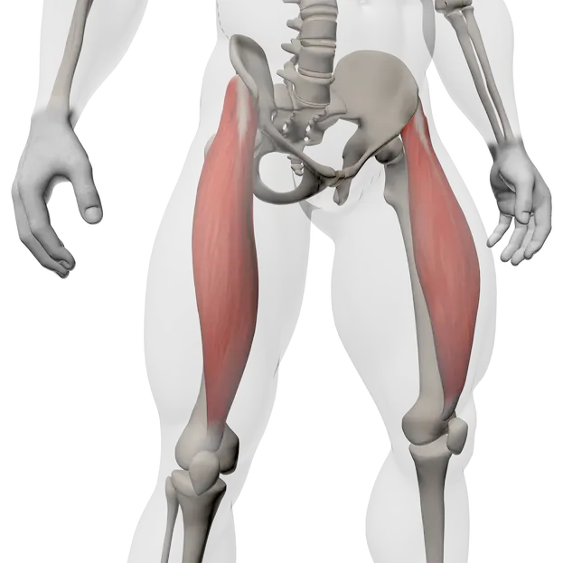
Rectus Femoris
Scientific name:
Rectus Femoris
Location:
Muscle located at the front of the thigh, in the center of the quadriceps, running from the pelvis to the knee.
Overall role:
Provides knee extension and hip flexion, with a key role in hip–knee coordination during dynamic movements.
Sports where this muscle is a key :
- Sprint / acceleration
- Football, rugby
- Jumping sports (athletics, basketball)
- Cycling
- Combat sports
- Team sports with frequent direction changes
Origin
- Anterior inferior iliac spine (AIIS)
- Upper border of the acetabulum (hip socket)
Insertion
- Tibial tuberosity via the quadriceps tendon and patellar ligament
Innervation
- Femoral nerve (roots L2–L4)
(nerve involved in knee extension and anterior thigh control)
Key points to remember
- Only bi-articular muscle of the quadriceps (acts on hip and knee)
- Highly active during explosive and repetitive actions
- Sensitive to neuromuscular fatigue
- Strong influence on pelvic position (anterior tilt if dominant)
Main actions
- Knee extension
- Hip flexion
Synergists (muscles working with it)
- Vastus medialis, vastus lateralis, vastus intermedius
- Iliopsoas (hip flexion)
- Tensor fasciae latae (anterior stability)
Antagonists (muscles with opposite action)
- Hamstrings (knee flexion)
- Gluteus maximus (hip extension)
Postural / stabilizing role
- Anterior pelvic stabilization during single-leg support
- Helps control the knee during braking and landing phases
- Acts as a force transfer muscle between hip and knee
- Engages early in movement but fatigues quickly if control is poor
Practical coaching interpretations:
- Weak → compensation by iliopsoas or lower back
- Overactive / tight → anterior pelvic tilt, hip or knee discomfort
- Reduced range → vastus dominance and loss of coordination
Activation test (bodyweight / light resistance)
- Goal: check rectus femoris activation without lumbar compensation.
- Setup:
- Standing, single-leg support.
- Free knee lifted to ~90°.
- Isometric hold for 10–15 seconds.
- What to observe:
- Clear tension at the front of the thigh.
- Stable pelvis, neutral spine.
- Interpretation:
- ➡️ Lower-back shaking → poor activation.
- ➡️ Mainly abdominal sensation → core compensation.
Weakness test (light load)
- Goal: detect specific strength loss.
- Setup:
- Single-leg leg extension.
- Light load, slow tempo (≈ 3–1–2).
- What to observe:
- Smooth control through the full range.
- Interpretation:
- ➡️ Speed increase at the end → concentric weakness.
- ➡️ Anterior hip discomfort → proximal overload.
Dominance / compensation test
- Goal: identify dominance of vasti or iliopsoas.
- Setup:
- Controlled step-up with high knee drive.
- What to observe:
- Fluid movement without excessive arching.
- Interpretation:
- ➡️ Excessive lumbar arch → iliopsoas / lumbar dominance.
- ➡️ Lateral thigh sensation → vastus lateralis dominance.
- Simple correction:
- Reduce range of motion.
- Increase isometric holds.
- Use slow tempo before adding load.
Activation / isolation
- Controlled leg extension (light load)
- Isometric knee lift holds
Functional / multi-joint
- Controlled step-ups
- Vertical split squats
- Technical high-knee drills (low volume)
Useful variations
- Slow unilateral work to limit lumbar compensation
Typical imbalances
- Rectus femoris dominance over vasti
- Quadriceps / hamstring imbalance
Related risks / pain
- Anterior knee pain
- Anterior hip discomfort
Warning signs
- Front thigh tightness
- Rapid fatigue during sprinting or stair climbing
- Excessive lower-back arch during effort
- Heavy leg extensions without control
- Explosive work with poor pelvic stability
- Excessive hip flexion ranges
- Repeated lumbar compensation patterns
- Controlled pelvic tilts in standing position
- Active hip mobility (slow knee lifts with gentle rotations)
- Slow, mindful walking to rebalance hip–knee coordination