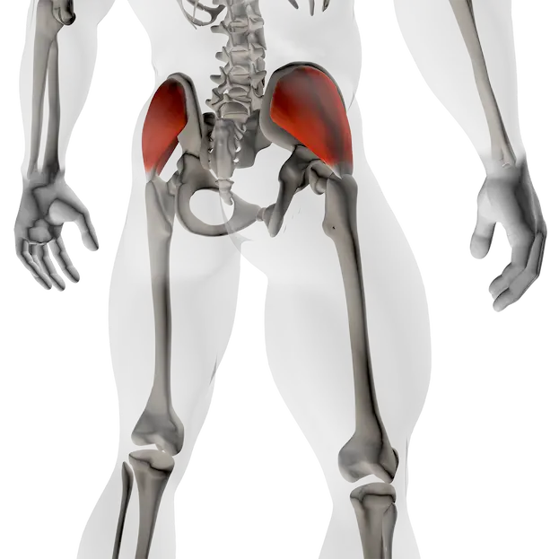
Gluteus Medius
Scientific name:
Gluteus Medius
Location:
Muscle located on the side of the hip, between the iliac bone and the upper femur.
Overall role:
Stabilises the pelvis and controls hip alignment, especially during single-leg support.
Sports where this muscle is a key :
- Running
- Fast walking / hiking
- Team sports with frequent single-leg support (football, basketball, handball)
- Combat sports
- Functional fitness / cross-training
Origin
- Outer surface of the ilium, between the anterior and posterior gluteal lines.
Insertion
- Lateral surface of the greater trochanter of the femur.
Innervation
- Superior gluteal nerve (L4–L5–S1)
- Key nerve for lateral pelvic stability.
Key points to remember
- Deep, postural muscle, not primarily aesthetic.
- Works continuously with the gluteus minimus.
- Often active in isometric control rather than visible movement.
Main actions
- Hip abduction (moving the leg away from the midline).
- Pelvic stabilisation during single-leg stance.
- Assists internal or external hip rotation, depending on joint position.
Synergists (muscles working with it)
- Gluteus minimus (fine pelvic control)
- Gluteus maximus (global hip control)
- Quadriceps (knee–hip alignment)
- Core muscles (lateral chain stability)
Antagonists (muscles with opposite action)
- Hip adductors
Postural / stabilizing role
- Prevents pelvic drop on the opposite side during walking or running.
- Maintains alignment between ankle, knee and hip.
- Acts as a lateral stabilising cable during single-leg loading.
- Transfers and absorbs forces between the ground and the pelvis.
Coaching-oriented interpretation:
- Weak → compensation through lower back, TFL, or adductors.
- Overactive / tight → lateral hip discomfort, reduced movement fluidity.
- Altered range of motion → excessive quadriceps or fascia lata involvement.
A poorly functioning gluteus medius quickly affects the knee, pelvis and lower back.
Activation test
Objective: check immediate gluteus medius activation.
Setup:
- Single-leg stance.
- Opposite foot lightly off the ground.
- Pelvis level, torso upright.
What to observe:
- Clear lateral hip engagement on the stance leg.
- No pelvic drop.
Interpretation:
- ➡️ Pelvic drop → insufficient activation.
- ➡️ Lower-back tension → compensation.
Weakness test
Objective: assess lateral control capacity.
Setup:
- Lateral band walk with a mini-band above the knees.
- Slow, controlled steps.
What to observe:
- Knee stays aligned.
- Pelvis remains stable.
Interpretation:
- ➡️ Knee collapsing inward → gluteus medius weakness.
- ➡️ Rapid loss of control → low endurance.
Dominance / compensation test
Objective: identify dominant compensators.
Setup:
- Shallow single-leg squat.
What to observe:
- Main area of effort sensation.
Interpretation:
- ➡️ Mostly quadriceps → anterior dominance.
- ➡️ Strong TFL tension → lateral compensation.
Simple correction:
- Reduce range of motion.
- Slow tempo.
- Short isometric holds.
Activation / isolation
- Single-leg hip abduction
- Single-leg balance with pelvic control
- Side leg raises with slow tempo
Functional / multi-joint
- Controlled single-leg squat
- Lateral lunge
- Step-up with controlled top position
Useful variations
- Extended isometric holds in single-leg stance
- Mini-band work for proprioceptive feedback
Typical imbalances
- Adductor or TFL dominance.
- Poor postural endurance.
Related risks / pain
- Lateral hip pain.
- Knee pain linked to dynamic valgus.
- Lower-back overload.
Warning signs
- Visible pelvic drop during walking.
- Instability on one leg.
- Rapid fatigue in unilateral stance.
- Excessive loading during hip abduction.
- Fast, uncontrolled movements.
- Large ranges of motion with loss of alignment.
- Lower-back or pelvic compensation.
- Controlled hip circles, progressive range.
- Slow lateral weight shifts with wide stance.
- Standing hip rotations, pelvis stable, calm breathing.
Goal: restore fluidity without deactivating, maintain motor control and joint awareness.